About Sinaps
We are grateful for the support of the Brain Initiative NIH funding in the project “Novel optrodes for large-scale electrophysiology and site-specific stimualtion” (U01NS094190-1) and to our project partners at IIT and Harvard University.
For the development of ChroMOS probes, we are also grateful to the European Commission for the support through the Marie Skłodowska-Curie Actions in the project “Toward monolithic CMOS based neural probes for stable chronic recording and brain machine interfaces” (ChroMOS project, H2020-MSCA-IF-2019, GA 896996).
About
Publications
Channels, Layout and Size Scalability of Implantable CMOS-Based Multielectrode Array Probes
Berdondini, L., et al.
69th IEEE International Electron Devices Meeting (2022), Invited Keynote
Publications
The SiNAPS Concept
Advantages
-
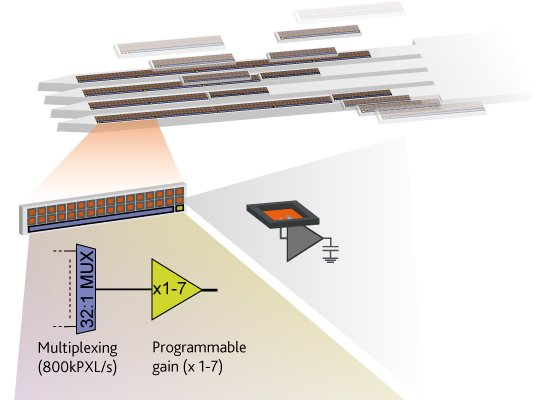
Modular circuit architecture
rapid design of probes with different layouts (single-/multi-shaft) -
Versatile fabrication flow
low-cost prototyping of CMOS-probes -
Compact and scalable devices
front-end circuits in-pixel avoid needs of large probe bases and allow >80 channels/mm2 of total device area.
Modular circuit architecture
different CMOS-probe layouts for in vivo electrophysiology
Active electrode-pixel
amplifiers and filters underneath each electrode
Sub-millisecond read-out circuits
On-probe addressing and time-division multiplexing circuits for whole array read-outs of thousands of electrodes at 25 ksample/s
Concept
Technical Specifications
One architecture, multiple layouts
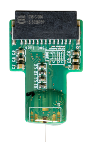
Single Shank
Amplifier | In-pixel (DC-input)
|
Eletrode Size | 20 x 20 µm² |
Eletrode Pitch | 28 µm |
Sampling Frequency | up to 25 kHz/channel |
Shank (L•W•T) | 6.5 • 10³ x 120 x 30 µm³ |
Base (L•W) | 1.7 x 1.12 mm² |
Number of shanks | 1 |
Shank’ Separation | NA |
Electrode/Channels | 512/512 |
RMS noise | 7.5 µV |
Power Consumption | <6 µW/electrode-pixel |
Electrode Material | Pt/Au |
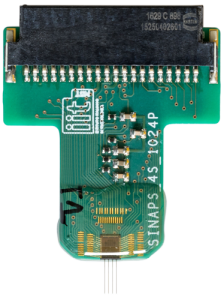
Multi Shank
Amplifier | In-pixel (DC-input)
|
Eletrode Size | 15 x 15 µm² |
Eletrode Pitch | 28 µm |
Sampling Frequency | up to 25 kHz/channel |
Shank (L•W•T) | 5 • 10³ x 80 x 30 µm³ |
Base (L•W) | 3.5 x 3 mm² |
Number of shanks | 4 |
Shank’ Separation | 560 µm |
Electrode/Channels | 1024/1024 |
RMS noise | 6.5 µV |
Power Consumption | <6 µW/electrode-pixel |
Electrode Material | Pt/Au |

ChroMOS
Amplifier | In-pixel (DC-input)
|
Eletrode Size | ø 10 µm |
Eletrode Pitch | 29.5 µm inside module and 90 µm between modules (modules of 8 electrodes) |
Sampling Frequency | up to 25 kHz/channel |
Shank (L•W•T) | 4 x 10³ x 26 x 20 µm³ |
Base (L•W) | 1 x 2 mm2 |
Number of shanks | 1 |
Shank’ Separation | N/A |
Electrode/Channels | 64/64 |
RMS noise | 6.5 µV |
Power Consumption | <6 µW/electrode-pixel |
Electrode Material | Pt/Au |
Recorded Data
Example of in vivo full-band neural recordings collected from SiNAPS single and multi-shank probes implanted in rodents' brain. The raw neural traces are filtered in the LFP (1-300 Hz) and AP (300-5000 Hz) frequency bands and spiking activity is subsequently isolated and extracted.
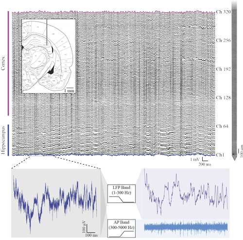

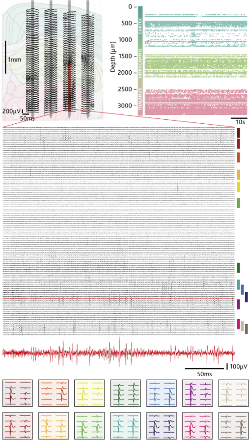
Thanks to A.Sirota (LMU) and A.Jackson (Newcastle University) for pilot experiments.
Contact
To get in touch with the SiNAPS team just fill in this form and soemone from the team will get back to you.